
From Munro Microabscess to Dermal Papillary Neutrophils: A Comprehensive Guide to the Histopathological Diagnosis of Psoriasis
Diagnosing atypical psoriasis requires more than clinical observation. This article summarizes Prof. Fang Sheng's presentation on the evolving histopathological features of psoriasis and key points for differential diagnosis.

Editor’s Note:
Psoriasis is a common chronic relapsing inflammatory skin disease affecting more than 60 million adults and children worldwide. In recent years, the use of biologic agents and small-molecule targeted therapies has led to breakthrough advances in the treatment of psoriasis, and accurate diagnosis is a prerequisite for standardized treatment. Typical psoriasis can be diagnosed on the basis of clinical manifestations alone; however, most atypical cases, including psoriasiform dermatitis, eczematous psoriasis, and other clinical variants, still require histopathological examination to establish the diagnosis. How to precisely identify the characteristic changes of psoriasis under the microscope and differentiate it from a variety of mimicking diseases is an important issue for dermatopathologists.
This article is based on the excellent presentation by Professor Fang Sheng from the Department of Dermatology, the First Affiliated Hospital of Chongqing Medical University, in the academic lecture "Histopathological Diagnosis and Differential Diagnosis of Atypical Psoriasis." It reviews the dynamic evolution of the histopathology of psoriasis, key diagnostic features, and differential diagnostic approaches, with the aim of providing a practical reference for clinicians.
1 Histopathological Evolution of Psoriasis at Different Stages of Development
The histopathological findings of psoriasis change dynamically with the stage of lesion development. Recognizing this evolutionary pattern can help avoid misdiagnosis caused by differences in the timing of biopsy.
Early lesions (less than 24 hours): At this stage, parakeratosis may be absent, and the granular layer is normal. The main changes are located in the dermal papillary layer, with dilated and congested capillaries, lymphocyte extravasation, mild perivascular lymphocytic infiltration, and tissue edema. Occasionally, lymphocytes extend into the epidermis with mild spongiosis. Such findings may resemble pityriasis rosea. However, if the clinical picture does not support pityriasis rosea, one should maintain an attitude of "psoriasis cannot be excluded" rather than readily ruling it out. Early psoriasis can be atypical; this is a diagnostic concept that must be established.
Papular stage: As the lesion progresses, focal parakeratosis appears, and neutrophils migrate through the epidermis and aggregate at the top of parakeratotic foci. Mitotic activity in the basal layer increases, with mild to moderate psoriasiform hyperplasia. The dermal papillary vessels are dilated and tortuous, and their lumina may contain neutrophils. At this stage, the granular layer is not yet thinned. A suggestive feature is the presence of neutrophils around blood vessels in the dermal papillary layer. The inflammatory infiltrate in psoriasis is usually predominantly lymphocytic, and the appearance of neutrophils in the dermal papillae supports the diagnosis of psoriasis. Guttate psoriasis may also show these changes, but epidermal hyperplasia is usually milder.
Plaque stage: This is the stage with the greatest diagnostic value for psoriasis. Parakeratotic areas gradually become confluent, and sometimes multiple layers of parakeratosis containing neutrophils alternate with orthokeratotic layers. Neutrophil aggregates within the stratum corneum (Munro microabscesses) are relatively common, whereas similar aggregates in the spinous layer (Kogoj microabscesses) are less common. The epidermis shows regular psoriasiform hyperplasia, with thinning of the suprapapillary epidermal plates. The dermal infiltrate contains activated T cells and a small number of neutrophils. At this stage, neutrophil aggregates can still be seen in the dermal papillae. Neutrophils migrate from the capillaries of the dermal papillae toward the epidermis and ultimately reach the stratum corneum to form microabscesses. This process explains why neutrophils may already appear in the dermal papillae before epidermal changes develop in early lesions.
Late lesions: Long-term scratching may superimpose changes of lichen simplex chronicus on psoriatic plaques, making differentiation difficult. Histopathologically, there may be three scenarios: ① psoriasis; ② psoriasis with superimposed lichen simplex chronicus; ③ lichen simplex chronicus . The key distinguishing features are as follows: in pure lichen simplex chronicus, the dermal papillae should show collagen sclerosis and irregular epidermal hyperplasia, whereas in cases with an underlying psoriatic component, the dermal papillae tend to show edema rather than sclerosis, and the granular layer is thinned. It is necessary to combine the clinical history and search for "clues" of psoriasis in the histopathology. Diagnostic reports may use cautious wording such as "favored diagnosis" or "cannot be excluded."
Resolving or treated lesions: The inflammatory infiltrate gradually decreases, the degree of epidermal hyperplasia diminishes, and the granular layer gradually recovers. At this time, the blood vessels in the dermal papillary layer may remain dilated, and the number of fibroblasts in this area increases with mild fibrosis. It usually takes 10–14 weeks of treatment for the histological findings to return completely to normal.
2 Interpretation of Key Histopathological Features
Munro microabscess and Kogoj microabscess: Neutrophil aggregates within parakeratotic areas of the stratum corneum are called Munro microabscesses, whereas those located in the granular layer or upper spinous layer are called Kogoj microabscesses. Both are commonly seen in psoriasis but can also be found in other skin diseases. The absence of microabscesses does not exclude psoriasis; a comprehensive judgment should be made in combination with scoring models and lesion maturity. When microabscesses are present, one should assess whether other psoriatic features are present and whether there is prominent spongiosis, exudation, infection, or eosinophilic infiltration. The simultaneous presence of Munro and Kogoj microabscesses is more diagnostically specific.
Eosinophils and spongiosis: Mild epidermal spongiosis and a small number of eosinophils are not uncommon in psoriasis. Psoriasis may show several or even more eosinophils, possibly as a secondary phenomenon or related to scratching or an allergic reaction. Therefore, psoriasis cannot be excluded solely on the basis of eosinophils. By comparison, intraepidermal serous crust is more specific than spongiosis. If more than 10 eosinophils are present or intraepidermal vesiculation develops, the reliability of a diagnosis of psoriasis should be reconsidered.
Pattern of inflammatory infiltration: Psoriasis does not show vacuolar alteration of the basal cell layer, which can help distinguish it from some mimickers. The depth of the inflammatory infiltrate also has some differential diagnostic significance: the dermal inflammatory infiltrate in psoriasis is usually concentrated in the dermal papillary layer, whereas other diseases may show deeper or more diffuse infiltration. Neutrophil aggregates around the capillaries of the dermal papillae suggest psoriasis.
Dermal papillary changes: These include capillary dilation, papillary edema, intrapapillary neutrophil aggregates, and papillary fibrosis, all of which are important auxiliary diagnostic clues for psoriasis.
Epidermal keratinization abnormalities: Reduction of the granular layer and confluent parakeratosis are common but lack specificity. Extensive reduction of the granular layer has some diagnostic significance.
Exploration of scoring models: Some investigators have attempted to construct a scoring model for plaque psoriasis, incorporating factors such as serous crust (with scoring/weighting if serous crust is absent), reduction or loss of the granular layer, psoriasiform hyperplasia, neutrophil aggregates, dermal neutrophils, dermal eosinophils, dermal papillary edema, and the depth of inflammatory infiltration. These factors were selected and assigned thresholds through regression analysis. The model is still in the exploratory stage, and clinical application should be cautious.
3 Differential Diagnosis Case Sharing
The histopathological differential diagnosis of psoriasis requires integration of clinical information and systematic analysis from multiple dimensions. First, determine the stage of the lesion: early lesions have limited diagnostic value, whereas plaque-stage features are the most typical. Second, pay attention to the presence and distribution of microabscesses; the coexistence of Munro and Kogoj microabscesses has relatively high specificity. Third, assess the degree of spongiosis and the number of eosinophils; marked spongiosis or numerous eosinophils should prompt reconsideration of the diagnosis. In addition, the depth and pattern of the inflammatory infiltrate, changes in dermal papillary blood vessels, and the regularity of epidermal hyperplasia are all important differential diagnostic points. For late or treated lesions, one should be aware that secondary changes may obscure the original features of psoriasis, and biopsy at multiple sites or follow-up re-examination may be considered when necessary.
4 The following are several case presentations:
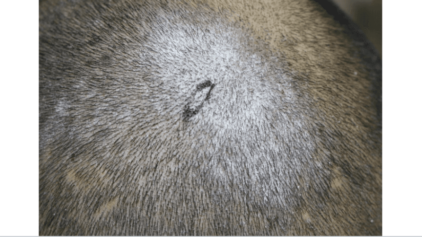
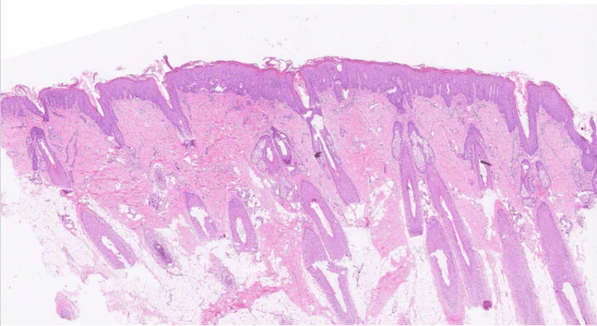
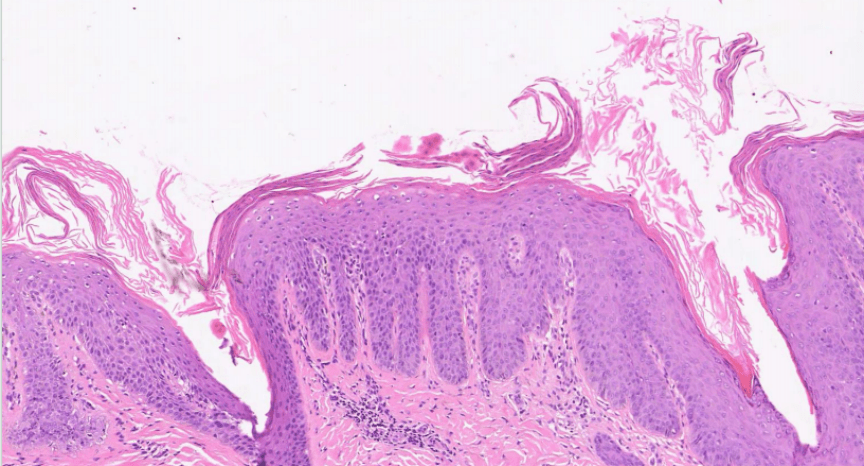
Fig. Seborrheic dermatitis
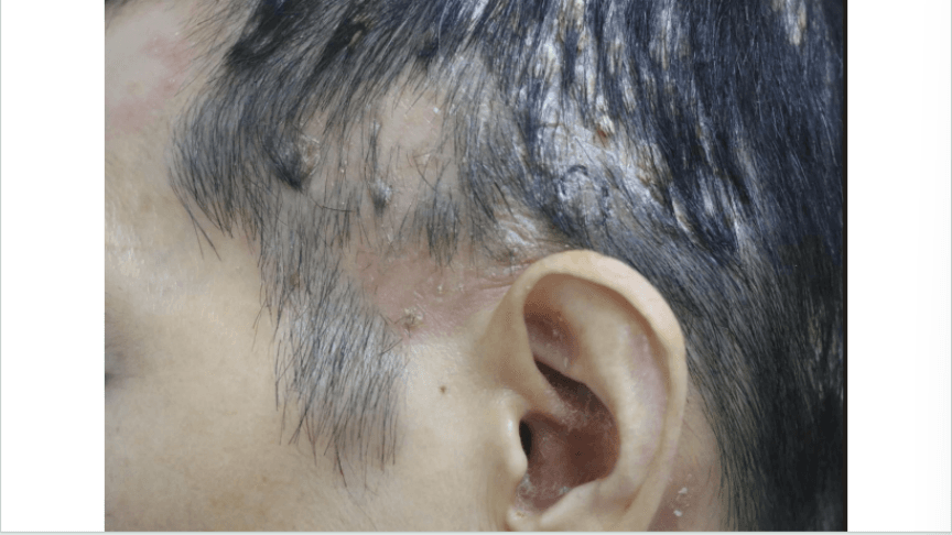
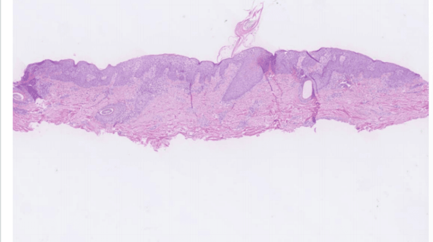
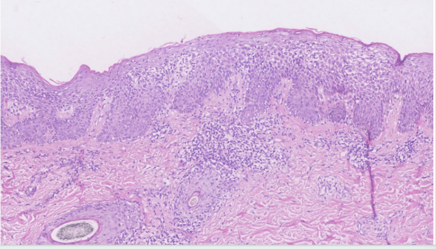
Fig. Eczema
5 Conclusion
The histopathological diagnosis of psoriasis is a dynamic and comprehensive judgment process. It requires pathologists to fully understand the pathological evolution of lesions at different stages, accurately grasp the diagnostic value and limitations of key histopathological features, and conduct systematic differential diagnostic analysis in combination with clinical information. With the advent of the biologic era, the treatment of psoriasis has achieved revolutionary progress, but precise diagnosis remains the cornerstone of effective treatment. For cases with atypical clinical manifestations or atypical pathological changes, histopathological examination remains an indispensable diagnostic tool. By systematically mastering the histopathological features of psoriasis and its differential diagnostic points, and by combining quantitative tools such as scoring models, diagnostic accuracy can be significantly improved, laying a foundation for precision treatment of patients.
Expert Profiles

Professor Fang Sheng
The First Affiliated Hospital of Chongqing Medical University
Specializes in the diagnosis and treatment of skin tumors, complex dermatoses, and immune-mediated skin diseases (such as psoriasis, pemphigus, scleroderma, etc.).